Players in the Pathogenesis of the Retroviral Infections

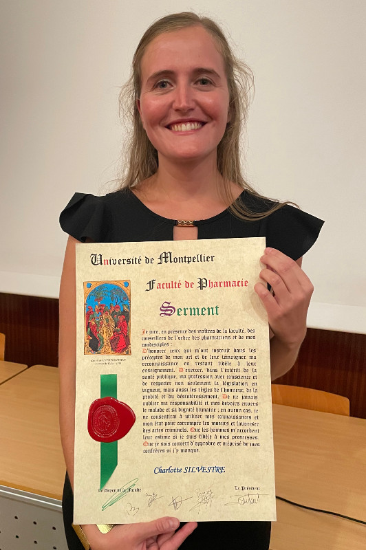
The team in May 2022. De gauche à droite : N Chazal, V Hebmann, JM Mesnard, C Mourouvin, L Espert, M Jansen, JM Peloponese, MA Houmey, J Tram, M Abrantes, B Beaumelle, B Pradel, N Audemard, C Silvestre, L Marty, A Gross.
Topics
Group 1: Roles of HBZ in HTLV-1-induced leukemia (Leader: Jean-Marie Peloponese)
Group 2: ASP, the antisens protein of HIV-1: Evolution, Immunological, Cellular and Viral impacts (Leader: Antoine Gross)
Group 3: Role of extracellular Tat during AIDS (Leader: Bruno Beaumelle)
Group 1. Roles of HBZ in HTLV-1-induced Leukemia
Human T-cell leukemia virus type 1 (HTLV-1) is the first retrovirus found to induce diseases in human. HTLV-1 causes an aggressive neoplastic disease, adult T-cell leukemia (ATL), neurodegenerative diseases, such as HTLV-1 associated myelopathy/tropical spastic paraparesis (TSP/HAM) and inflammatory diseases such as uveitis. HTLV-1 infects around 15 million people in the world and is mainly endemic of the intertropical region like South America, Caribbean island, Africa and south japan. HTLV-1 is a complex retrovirus, which encodes regulatory genes (tax and rex) and several accessory genes, such as p30, p12, p13 and HTLV-1 bZIP factor (HBZ). Among them, tax is thought to play a central role in transformation of infected cells. However, since it is a major target of cytotoxic T-lymphocytes, its expression is often silenced in ATL cells to escape the host immune system. The HBZ gene, first characterized in our laboratory in 2002, is encoded by the minus strand of HTLV-1, and it contains a basic leucine zipper domain. Interestingly enough, while tax is repressed in infected cells, transcription of the HBZ gene was detected in all of the ATL cell lines and primary ATL cases. We have shown that HBZ is able to sustain its own expression in infected cells via a regulation loop with JunD and SP1. We have also shown that HBZ is able to transform cells in vitro.
Our research on cellular and molecular mechanisms in the HBZ-induced pathogenesis are currently focusing on 2 Axes:
– We are investigating the importance of the interplays between HBZ and AP-1 pathway in HTLV-1 infected cells.
– In collaboration with clinicians, we are trying to understand the role of HBZ in the chemoresistance of ATL cells and to develop new therapeutic approaches against HBZ.
Group 2. ASP, the antisense protein of HIV-1 : Evolution, Immunology, Cellular, and Viral impacts
Our research project focuses on the HIV-1 ASP protein (AntiSense Protein). Using several experimental approaches (virology, cell biology, immunology, bioinformatics, studies in HIV-1 infected patients), our project aims to determine the function of this protein and to understand its role in the pathogenesis of AIDS (viral latency, chronicity). HIV-1 produces its proteins from the integrated proviral DNA (consisting of two LTR5′ and one LTR3′) through transcriptional activity of a promoter located in the LTR-5′ (Long Terminal Repeat). However, like HTLV-1 which has a transcriptional activity leading to the production of the HBZ protein (see HBZ project), we have shown that the HIV-1 LTR-3′ also has an antisense transcriptional activity allowing the production of the ASP protein.

It was first predicted in 1988, that there may be an Open Reading Frame (ORF) on the negative strand of the Human Immunodeficiency Virus type 1 (HIV-1) genome that could encode a protein named AntiSense Protein (ASP) (Savoret et al., Frontiers in Microbiol, 2021). Despite early skepticism and the lack of specific tools to selectively identify rare antisense transcripts and detect a strongly hydrophobic protein like ASP, the presence of antisense transcripts was observed (Landry et al., Retrovirology, 2007) as well as the presence of CD8+ T cells directed against several ASP peptides in HIV-1-infected patients (Bet et al., Retrovirology, 2015). Recently, the presence of ASP-specific antibodies was detected in the plasma of HIV-1-infected individuals (Savoret et al., 2020) further suggesting that ASP is expressed and immunogenic in vivo. Finally, using an evolutionary study developed in collaboration with LIRMM informaticians and performed on more than 23,000 sequences, we demonstrated that the ASP gene emerged during the emergence of the pandemic HIV-1 in the early twentieth century (Cassan et al., PNAS, 2016).

Our current research focuses on:
– The study of ASP function and its relationship with infection and immune response.
– The link between ASP and the immune status of the HIV_1 infected patient under antiretroviral treatment.
– The evolutionary mechanisms of ASP in HIV-1.
COVID-19: More recently, we have initiated studies on SARS-CoV-2, in particular on some poorly studied proteins of the virus by focusing on cell biology aspects and antibody response in patients.
Grants:
– Fédération Hospitalo-Universitaires Infection Chronique (FHU Inch): 2020-2023: Doctoral fellowship(3 years) – collaboration with Dr. Alain Mackinson (at infectious and tropical diseases department at Montpellier Hospital)
– Sidaction: 2020-2023 : Doctoral fellowship (3 years)
– Sidaction: 2019: Doctoral fellowship (1 year) for the fourth year thesis
– “Direction des relations avec les entreprises du CNRS”: 2019-2020
– UM/CHU: 2015-2018: Doctoral fellowship (3 years)
– “CNRS mission Interdisciplinarité” 2013-2016
Group 3. Role of extracellular HIV-1 Tat during AIDS
Previous work
Our group studies the intracellular and transcellular transport of HIV-1 Tat protein. This protein is well known and studied because it is required for the transcription of viral genes. Its concentration and activation level regulate cell exit from latency. We showed that Tat is secreted using an unconventionnal pathway based on Tat binding to PI(4,5)P2 on the inner leaflet of the plasma membrane (Rayne et al, 2010). Circulating Tat can then interact with a number of cell types using receptors such as the LRP. We found that Tat is then endocytosed using the clathrin/AP-2 pathway (Vendeville et al, 2004). Once in endosomes, low pH triggers a conformational change inducing Tat membrane insertion. Tat single Trp (Trp11) is required for this insertion process (Yezid et al, 2009). Tat translocation to the cytosol is catalyzed by Hsp90, a cytosolic chaperone (Vendeville et al, 2004). Once in the cytosol Tat induces a number of cell responses, for instance the secretion of pro-inflammatory cytokines by monocytes (Rayne et al, 2004). Incoming Tat also binds to PI(4,5)P2. Recombinant Tat shows an affinity for this phosphoinositide that is much higher than that of cell proteins because Tat inserts the sidechain of its Trp in the membrane upon PI(4,5)P2 binding (Debaisieux et al, 2012). In uninfected cells, Tat is palmitoylated and this modification will further stabilize its membrane association and inhibit secretion. Tat palmitoylation requires the prolylisomerase cyclophilin A (CypA). Tat is not palmitoylated in infected cells because CypA is encapsidated by HIV-1 (~200 CypA/virion), thereby clearing the cell of CypA and enabling efficient Tat secretion by infected cells (Chopard et al, 2018) (Fig.2). In uninfected cells, Tat palmitoylation makes Tat resident on PI(4,5)P2, thereby severely perturbating the assembly and function of different cell machineries using this phosphoinositide. We accordingly showed that circulating Tat prevents the recruitment of annexin 2 and cdc42, thereby perturbating neurosecretion (collaboration with the group of Nicolas Vitale, INCI, Strasbourg (Tryoen-Toth et al, 2013)) and phagocytosis (Debaisieux et al, 2015), respectively (Fig.1). Tat also affects the function of ionic channels in cardiomyocytes (collaboration with the group of Gildas Loussouarn, INSERM, Nantes) (Es-Salah-Lamoureux et al, 2016). It should be noted that HIV patients indeed show these dysfunctions.
Our results are summarized in Figs. 1 et 2. These projects were funded by the ANRS, Sidaction and FRM and performed by Fabienne Rayne (PostDoc), Agnés Vendeville, Hocine Yezid, Solène Debaisieux, Christophe Chopard, Bao Viet Tong et Malvina Schatz (PhDs).


*Present work
We are interested in the effect of Tat on the multiplication of opportunistic pathogens following HIV infection (collaborations Laurent Kremer and Laura Picas from IRIM, Oliver Neyrolles and Christel Verollet from IPBS Toulouse), in a potential mechanism of Tat encapsidation (collaboration Mickaël Blaise and Laurent Chaloin from IRIM, Pierre-Emmanuel Milhiet and Jean-François Guichou, CBS Montpellier) and in the development of new HIV latency reversing agents (collaboration L. Chaloin), molecules that were just patented. These projects are funded by Sidaction and SATT AXLR.
At a glance
The team is interested in viral proteins whose involvement in infection (HIV-1 Tat) or cell transformation (HBZ, the Human bZIP factor of HTLV-1) are well established, but also to a viral protein whose function is still unknown (the HIV-1-antisense protein (ASP)). Our studies aim to identify the function and the biological activity of these proteins both in viral multiplication and in the pathogenic effects linked to infection. Furthermore, we study the cellular answer to viral infections, in particular the autophagy process.
Funding
