THE PROJECTS
Mycobacterium tuberculosis, the etiologic agent of tuberculosis, represents a major cause of death worldwide due to a unique infectious agent. Since the mid-80s, there has been a prominent progression of the disease, substantiated by the spread of the HIV pandemic and the emergence of multidrug-resistant M. tuberculosis strains. In addition, atypical mycobacteria, including Mycobacterium abscessus, represent an emerging health problem in industrialized countries, and are notorious for being highly resistant to most antibiotic treatments. Thus, there is an urgent need to develop new therapies to combat these infections. One key aspect characterizing pathogenic mycobacteria resides in their capacity to persist within the phagocytic cells for several years/decades, which is strongly associated with the presence of a very unusual cell envelope. These cell wall components play a key role in driving host-pathogen interactions necessary for the establishment and persistence of the infection and represent valid targets for several antitubercular drugs.
In this context, we explore the mycobacterial cell envelope to decipher its role in the physiopathological events characterizing the infection and to identify new pharmacological targets. Our work focuses on major cell wall (glycol)lipidic components with respect to their biosynthesis, regulation, and contribution/role in virulence in pathogenic mycobacteria.
Our research program aims to investigate virulence factors in Mycobacterium tuberculosis as well as in non-tuberculous mycobacteria, such as M. abscessus, by studying genes involved in biosynthesis, catabolism and transport of mycobacterial cell wall components. Among these components are mycolic acids (long-chain fatty acids) and a vast array of lipids and glycolipids sharing exotic structures and participating in the immunopathology of the infection. Besides determining the metabolic pathways and the mechanisms controlling expression of these molecules, we are also interested in elucidating the structures of the (glycol)lipid components and their biological functions to better define their role in the physiopathology of the infection and to discover new targets of pharmacological interest. The molecular mechanisms responsible for virulence and physiopathology of atypical mycobacteria remaining elusive, we also develop alternative models of infection to apprehend and describe new mechanisms of immune evasion and persistence in the infected host.
Our studies can be summarized by 3 mains interconnected goals.
Aim 1: Microbiology of tuberculous and non-tuberculous mycobacteria.
We focus essentially on the biosynthetic and catabolic pathways of lipidic and glycolipidic cell wall associated components.
Genetic, biochemical and crystallographic studies are employed to discover and characterize enzymes involved in the biosynthesis, transport and remodeling of the mycobacterial cell wall. The objective here consists of understanding the role of components of the envelope in the adaptation of pathogenic mycobacteria to their environment and to establish a successful infection process within the host as well as to identify and validate new targets of therapeutic interest to fight against mycobacterial infections. This aim includes also the elucidation of structures and biological functions of glycolipids in atypical mycobacteria (M. marinum and M. abscessus complex) and to determine their contribution in the the pro-inflammatory response and granuloma formation.
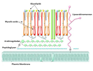
Schematic structure of the mycobacterial envelope. Besides a plasmic membrane, the cell wall consists of a complex network of proteins, lipoglycans and (glyco)lipids, which participate directly in the interaction with the host cells. Because of their uniqueness in Mycobacterium, the enzymes involved in the biosynthesis of the cell wall components represent attractive targets for future drug development.
Aim 2: Mode of action of antimycobacterial drugs.
We study the mechanisms of activation and action of molecules inhibiting the biosynthesis of cell wall components, with a special emphasis on mycolic acids.
This aim consists essentially of determining the molecular targets and mode of action of several antimycobacterial drugs, of synthesizing structural analogues that are more efficient and less toxic than the parental molecules and to evaluate the anti-mycobacterial properties of these compounds in vitro and in vivo in a zebrafish model of infection (see Aim 3).
Another key aspect of this axis consists to describe the mechanisms responsible for the intrinsic resistance of atypical mycobacteria to most antitubercular drugs and to discover new active molecules against these species. This implies the screening of chemical libraries, the selection of resistant strains to the selected active inhibitors and identification/characterization of the therapeutic targets through the combination of genetic/biochemical and crystallographic techniques.
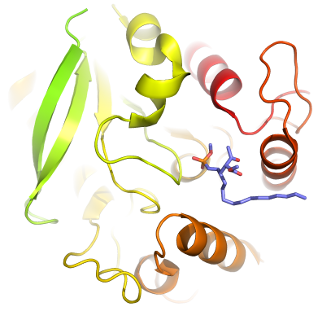
Three-dimensional structure of Ag85C bound to a potent inhibitor cyclophostin
Aim 3: Alternative models of infection to study mycobacterial virulence.
We develop amoeba and zebrafish (Danio rerio) models to identify new virulence genes and to image in real time the chronology of the infection.
The amoeba model is particularly adapted to the screening of transposon libraries to select for instance attenuated mutants of M. marinum or M. abscessus and to evaluate and compare their capacity of intracellular growth and survival. The zebrafish is also used to evaluate and compare virulence of mutants and, due to its optical transparency, this model is particularly suited to image the infection process at a spatiotemporal level. In this context, it has been successfully used to propose mycobacterial cords as a new mechanism of immune evasion by preventing mycobacteria to be phagocytosed by macrophages and neutrophils. In addition, embryos can be used to visualize, in real time, the pharmacological activity of molecules/drugs or to study the role of cell wall components in the induction of an inflammatory granulomatous response during infection with M. marinum and M. abscessus.
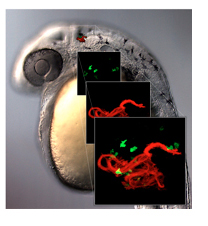
Zebrafish embryo infected with the rough variant of M. abscessus expressing mCherry (red). Microinjection was performed in the caudal vein in the mpx::GFP transgenic line harbouring green fluorescent neutrophils. The image shows the presence of a massive mycobacterial cord (red) surrounded by neuthrophils (green) in the brain.
POST-DOC, PhD and undergraduate students
We are looking for strong candidates (microbiologists, biochemists, structural biologists) as well as researchers or technicians/engineers with positions at CNRS, INSERM or University who are interested by our interdisciplinary approaches, combining basic and translational research in mycobacteriology.
Team Members
|
|

2026
Smooth to rough morphotype switching, a mechanism of phage resistance in Mycobacterium abscessus. J. H. Liew, A. Norman, M. Illouz, T.-H. Teo, R. Delli Ponti, C. Hamela, L. Mohan, R. Lew, C. Ron, K. E. Low, W. Daher, A. Chiam, J. Maw, S. Yan, R. Sorayah, S. H. Oehlers, B. W. Jhun, J. W. P. Teo, I. Bonne, R. Wintjens, K. Pethe, R. G. Huber, L. Kremer, and P. Bifani. 2026. Proc. Natl. Acad. Sci. USA. In Press.
Complete genome sequence of a multi-drug-resistant Mycobacterium avium subspecies hominissuis isolated from a patient with lung infection. F. Biet, C. Hamela, F. Roquet-Baneres, S. Melo, T. Cochard, S. Lameiras, S. Baulande, H. Chiapello, V. Loux, and L. Kremer. 2026. Microbiol. Resour. Announc. 5:e0001426.
Mycobacterium abscessus research: learning from challenges. R. Z. Treen, M. Gonzalez-Juarrero, M. Jackson, P. Lapierre, L. Kremer, P. Ghosh, A. K. Ojha. 2026. J. Bacteriol. 4:e0043625.
Isoniazid-derived vanillin and salicylaldehyde hydrazones: Promising antimycobacterials with KatG-dependent activation. N. Gupta, F. Roquet-Baneres, N. Henry, A. Anand, P. Roussel, L. Kremer, and V. Kumar. 2026. Bioorg. Chem. 173: 109644.
2025
TCS-Isatin-INH triads as potent and selective anti-mycobacterial with high efficacy against Mycobacterium tuberculosis. Shekhar, F. Roquet-Banères, L. Kremer, and V. Singh. 2025. Submitted to Eur. J. Med. Chem. Rep. 15: 100307.
EccC3 is essential for pathogenesis of rough Mycobacterium abscessus in zebrafish. Y. Tasrini, W. Daher, and L. Kremer. 2025. mSpectrum. 13: e0194925.
Iron-Sulfur proteins in mycobacteria: Master regulators of physiology and pathogenesis. V. Berdal, B. Py, S. Ollagnier de Choudens, L. Kremer, and W. Daher. 2025. Trends Microbiol. 16: S0966-842X(25)00205-7.
Isoniazid-Rhodanine molecular hybrids: Design, synthesis, antimycobacterial activity and computational validation. G. Kumar, S. Mishra, P. Seboletswe, N. Manhas, S. Ghumran, F. Roquet-Banères, L. Kremer, G. Bhargava, and P. Singh. 2025. RSC Adv. 15: 31272-31288.
Rationally designed InhA inhibitors: A comparative anti-tubercular activity study of sulfonate esters of isoniazid hydrazones and their structurally flexible benzyl analogues. M. G. Kadima, S. Mishra, G. Kumar, P. Seboletswe, F. Roquet-Banères, M. Foubert, L. Kremer, R. Karpoormath, and P. Singh. 2025. Chem. Biol. Drug Des. 106: e70171.
Rational design and antimycobacterial evaluation of aryl sulphonamide-linked isoniazid hydrazones against Mycobacterium tuberculosis. M. G. Kadima, S. Mishra, G. Kumar, P. Seboletswe, A. Kajee, Ankit, F. Roquet-Banères, M. Foubert, L. Kremer, R. Karpoormath, and P. Singh. 2025. ChemMedChem. 17: e202500398.
In vivo antimicrobial activity of engineered mesoporous silica nanoparticles targeting intracellular mycobacteria. J. J. Aguilera-Correa, Y. Tasrini, M. Gisbert-Garzarán, A. Boulay, T. Carvalho, F. P. Blanchet, M. Vallet-Regi, and L. Kremer. 2025. Nat. Comm. 16: 7388.
P3MA: A promising mycobacteriophage infecting Mycobacterium abscessus. A. Broncano-Lavado, J. J. Aguilera-Correa, F. Roquet-Banères, L. Kremer, A. Mediero, M. Seoane-Blanco, M. J. van Raaij, I. Pagán, J. Esteban, and M. García-Quintanilla. 2025. Antibiotics. 14: 801.
Mapping of mycobacterial enzymes involved in triacylglycerol accumulation as intrabacterial lipid inclusions using activity-based multi-target inhibitor probes. R. Avellan, J. Lehoux, T. Francis, T. Dargham, L. Celik, A. Guy, I. Poncin, V. Point, L. Kremer, T. Durand, S. Audebert, L. Camoin, C. D. Spilling, P. Santucci, C. Crauste, S. Canaan, and J.-F. Cavalier. 2025. ACS Infect. Dis. 11: 1589-1605
Genome Sequence of Mycobacterium abscessus phage P3MA. A. Broncano-Lavado, F. Roquet-Banères, L. Kremer, D. A. Russell, D. Jacobs-Sera, T. G. Reidy, J. J. Aguilera-Correa, J. Esteban, G. F. Hatfull and M. García-Quintanilla. 2025. Microbiol. Resour. Announc. 22: e0016925.
Zebrafish models for drug discovery and therapeutic validation against non-tuberculous mycobacteria. M. D. Johansen, C. Hamela, Y. Ding, and L. Kremer. 2025. Cold Spring Harb. Perspect. Med. 27: a041832.
Isoniazid-dihydropyrimidinone molecular hybrids: Design, synthesis, antitubercular activity and cytotoxicity investigations with computational validation. G. Kumar, P. Seboletswe, S. Mishra, N. Manhas, S. Ghumran, N. Kerru, F. Roquet-Banères, M. Foubert, L. Kremer, G. Bhargava, P. Singh. 2025. ChemMedChem. 11: e2400949
A glycosylated lipooctapeptide promotes uptake and growth of Mycobacterium abscessus in the host. L.-David Leclercq, V. Le Moigne, W. Daher, M. Cortes, B. Viljoen, Y. Tasrini, X. Trivelli, H. Lavanant, I. Schmitz-Afonso, N. Durand, F. Biet, Y. Guérardel*, L. Kremer*, and J.-L. Herrmann*. 2025. Nat. Comm. 16: 3326. *Co-last corresponding authors.
A proteomic and functional view of intrabacterial lipid inclusion biogenesis in mycobacteria. T. Dargham, J. J. Aguilera-Correa, R. Avellan, I. Mallick, L. Celik, G. Brasseur, I. Poncin, V. Point, S. Audebert, L. Camoin, W. Daher, J.-F. Cavalier, L. Kremer, and S. Canaan. 2025. mBio. 25: e0147524.
Deletion of ESX-3 and ESX-4 secretion systems in Mycobacterium abscessus results in highly impaired pathogenicity. W. Daher*, V. Le Moigne, Y. Tasrini, S. Parmar, D. L. Sexton, J. J. Aguilera-Correa, V. Berdal, E. I. Tocheva, J.-L. Herrmann, and L. Kremer*. 2025. Commun. Biol. 8: 166. *Corresponding authors.
SQ31f is a potent non-tuberculous mycobacteria antibiotic by targeting specifically the mycobacterial F-ATP synthase. P. Ragunathan, P. Sae-Lao, A. Harikishore, W. Daher, F. Roquet-Banères, L. Kremer, R. W. Bates, and G. Grüber. 2025. J. Antimicrob. Chemother. 80: 270-280.
2024
Design, synthesis, and anti-mycobacterial evaluation of Triclosan-Isoniazid hybrids. N. Gupta, S. Chowdhari, F. Roquet-Banères, L. Kremer, and V. Kumar. 2024. Chem. Biodiversity 13: e202401967.
Cyclophostin and Cyclipostins analogues counteract macrolide-induced resistance mediated by erm(41) in Mycobacterium abscessus. M. Sarrazin, I. Poncin, P. Fourquet, S. Audebert, L. Camoin, Y. Denis, P. Santucci, C. D. Spilling, L. Kremer, V. Le Moigne, J.‑Louis Herrmann, J.-F. Cavalier, and S. Canaan. 2024. J. Biomed. Sci. 31: 103.
Trehalose polyphleates participate in Mycobacterium abscessus fitness and pathogenesis.
S. Malmsheimer, W. Daher, Y. Tasrini, C. Hamela, J. J. Aguilera-Correa, C. Chalut, G. F. Hatfull and L. Kremer. 2024. mBio. 30: e0297024. DOI: 10.1128/mbio.02970-24.
A dTDP-L-Rhamnose 4-epimerase required for glycopeptidolipid biosynthesis in Mycobacterium abscessus.
J. J. Aguilera-Correa, F. Wei, L.-D. Leclercq, Y. Tasrini, E. Mullapudi, W. Daher, K. Nakajima, S. Canaan, J.-L. Herrmann, M. Wilmanns, Y. Guérardel, L. Wen, and L. Kremer. 2024. J. Biol. Chem. 300: 107852. DOI: 10.1016/j.jbc.2024.107852.
Combination of serological and cytokine release assays for improved diagnosis of childhood tuberculosis in Zambia (PROMISE-TB).
E. Tuaillon, M. Mwyia, K. Bollore, A. Pisoni, P.-A. Rubbo, M. Richard, L. Kremer, M. M.W. Tonga, D. M. Chanda, M. Peries, R. Vallo, S. Eymard-Duvernay, M. D’Ottavi, C. Kankasa, P. Van de Perre, J.-P. Moles, and N. Nagot. 2024. Int. J. Infect. Dis. 148: 107248. DOI: 10.1016/j.ijid.2024.107248.
Rational design and microwave-promoted synthesis of triclosan-based dimers: Targeting InhA for anti-mycobacterial profiling.
Shekhar, F. Roquet-Banères, A. Anand, L. Kremer, and V. Kumar. 2024. R. Soc. Open Sci. 11: 240676. DOI: 10.1098/rsos.240676.
The diversity of clinical Mycobacterium abscessus isolates in morphology, glycopeptidolipids and infection rates in a macrophage model.
V. Pichler, L. Dalkilic, G. Shoaib, T. Shapira, L. Rankine-Wilson, Y.-M. Boudehen, J. D. Chao, D. Sexton, M. Prieto, B. S. Quon, E. I. Tocheva, L. Kremer, W. Hsiao, and Yossef Av-Gay. 2024. J. Med. Microbiol. 73: 001869. DOI: 10.1099/jmm.0.001869.
Cu-promoted synthesis of triclosan-Mannich and Glaser adducts: anti-mycobacterial evaluation with in silico validations.
Shekhar, M. Alcaraz, A. Anand, R. K. Sharma, L. Kremer, and V. Kumar. 2024. Future Med. Chem. 16: 949-961. DOI: 10.4155/fmc-2023-0298.
Evaluation and activity of new porphyrin-peptide cage-type conjugates for the photoinactivation of Mycobacterium abscessus.
M. Alcaraz, S. Lyonnais, C. Ghosh, J. Aguilera-Correa, S. Richeter, S. Ulrich, and L. Kremer. 2024. mSpectrum. 12: e0000624. DOI: 10.1128/spectrum.00006-24.
Loss of LpqM proteins in Mycobacterium abscessus is associated with impaired intramacrophage survival.
Y.-M. Boudehen, W. Daher, F. Roquet-Baneres, and L. Kremer. 2024. mSpectrum. 12: e0383723. DOI: 10.1128/spectrum.03837-23.
High efficacy of the F-ATP synthase inhibitor TBAJ-5307 against non-tuberculous mycobacteria in vitro and in vivo.
P. Ragunathan, P. Sae-Lao, C. Hamela, M. Alcaraz, A. Krah, W. Han Poh, C. Jia Ern Pee, A. Yick Hou Lim, S. A. Rice, K. Pethe, P. John Bond, T. Dick, L. Kremer*, R. W. Bates*, and G. Grüber*. 2024. J. Biol. Chem. 300:105618. *Co-last corresponding authors.
Modeling non-tuberculous mycobacterial infections in zebrafish.
D. Johansen, H. P. Spaink, S. H. Oehlers, and L. Kremer. 2024. Trends Microbiol. S0966-842X(23)00329-3.
2023
Mycobacterium abscessus, a complex of three fast-growing subspecies sharing virulence traits with slow-growing mycobacteria.
Lagune, L. Kremer, and J.-L. Herrmann. 2023. Clin. Microbiol. Infect. S1198-743X(23)00485-8.
Tailoring selective triclosan azo-adducts: Design, synthesis, and anti-mycobacterial evaluation.
Shekhar, M. Alcaraz, P. Seboletswe, N. Manhas, L. Kremer, P. Singh, and V. Kumar. 2023. Heliyon. 9: e22182.
Intrabacterial lipid inclusion-associated proteins: A core machinery conserved from saprophyte Actinobacteria to the human pathogen Mycobacterium tuberculosis.
Dargham, I. Mallick1, L. Kremer, P. Santucci, and S. Canaan. 2023. FEBS Open Bio. 13: 2306-2323.
Silencing essential gene expression in Mycobacterium abscessus during infection.
Y.-M. Boudehen, Y. Tasrini, J. J. Aguilera-Correa, M. Alcaraz, and L. Kremer. 2023. mSpectrum. 13: e0283623.
Characterization of Mycobacterium abscessus colony-biofilms based on bi-dimensional images.
J. J. Aguilera-Correa, Y.-M. Boudehen, and L. Kremer. 2023. Antimicrob. Agents Chemother. 67: e0040223.
Therapeutically useful mycobacteriophages BPs and Muddy require trehalose polyphleates.
K. S. Wetzel, M. Illouz, L. Abad, H. G. Aull, D. A. Russell, R. Garlena, M. Cristinziano, S. Malmsheimer, C. Chalut, G. F. Hatfull*, and L. Kremer*. 2023. Nat. Microbiol. 8: 1717-1731 *Co-last corresponding authors.
Multiple Mycobacterium abscessus O-acetyltransferases influence glycopeptidolipid structure and colony morphotype.
M. Illouz, L.-D. Leclercq, C. Dessenne, G. Hatfull, W. Daher, L. Kremer*, and Y. Guérardel*. 2023. J. Biol. Chem. 299: 104979. *Co-last corresponding authors.
New therapeutic strategies for Mycobacterium abscessus pulmonary diseases – Untapping the mycolic acid pathway.
M. Alcaraz, T. E. Edwards, and L. Kremer. 2023. Expert Rev. Anti. Infect. Ther. 21: 813-829.
In vitro and in vivo efficacy of NITD-916 against Mycobacterium fortuitum.
F. Roquet-Banères, M. Alcaraz, C. Hamela, J. Abendroth, T. E. Edwards, and L. Kremer. 2023. Antimicrob. Agents Chemother. e01607-22.
Mycobacterial biotin biosynthesis counters airway alkalinity. W. Daher, and L. Kremer. 2023. Nat. Microbiol. 8: 369-370.
Mycobacterium abscessus alkylhydroperoxide reductase C promotes cell invasion by binding to tetraspanin CD81. J. Karam, F. P. Blanchet, E. Vivès, P. Boisguérin, Y.-M. Boudehen, L. Kremer*, and W. Daher*. 2023. iScience. 26, 106042. *Co-last corresponding authors.
Human IRF1 governs macrophagic IFN-γ immunity to mycobacteria.
J. Rosain, A.-L. Neehus, J. Manry, R. Yang, J. Le Pen, W. Daher, Z. Liu, Y.-H. Chan, N. Tahuil, Ö. Türel, M. Bourgey, M. Ogishi, J.-M. Doisne, H. M. Izquierdo, T. Shirasaki, T. Le Voyer, A. Guérin, P. Bastard, M. Moncada-Velez, J. E. Han, T. Khan, F. Rapaport, S.-H. Hong, A. Cheung, K. Haake, B. C. Mindt, L. Perez, Q. Philippot, D. Lee, P. Zhang, D. Rinchai, F. Al Ali, M. M. A. Ata, M. Rahman, J. N. Peel, S. Heissel, H. Molina, Y. Kendir-Demirkol, R. Bailey, S. Zhao, J. Bohlen, M. Mancini, Y. Seeleuthner, M. Roelens, L. Lorenzo, C. Soudée, M. E. J. Paz, M. L. Gonzalez, M. Jeljeli, J. Soulier, S. Romana, A.-S. L’Honneur, M. Materna, R. Martínez-Barricarte, M. Pochon, C. Oleaga-Quintas, A. Michev, M. Migaud, R. Lévy, M.-A. Alyanakian, F. Rozenberg, C. A. Croft, G. Vogt, J.-F. Emile, L. Kremer, C. S. Ma, J. H. Fritz, S. Lemon, A. N. Spaan, N. Manel, L. Abel, M. R. MacDonald, S. Boisson-Dupuis, N. Marr, S. G. Tangye, J. P. Di Santo, Q. Zhang, S.-Y. Zhang, C. M. Rice, V. Béziat, N. Lachmann, D. Langlais, J.-L. Casanova, P. Gros, and J. Bustamante. 2023. Cell. 186: 1-25.
Synthesis and testing of analogs of the tuberculosis drug SQ109 against bacteria and protozoa: Identification of lead compounds against Mycobacterium abscessus and malaria parasites.
M. Stampolaki, S. R. Malwal, N. Alvarez-Cabrera, Z. Gao, M. Moniruzzaman, S. O. Babii, N. Naziris, A. Rey-Cibati, M. Valladares-Delgado, A. L. Turcu, K.-H. Baek, T.-N. Phan, H. Lee, M. Alcaraz, S. Watson, M. van der Watt, D. Coertzen, N. Efstathiou, M. Chountoulesi, C. M. Shoen, Ioannis P. Papanastasiou, J. Brea, M. H. Cynamon, L.-M. Birkholtz, L. Kremer, J. H. No, S. Vázquez, G. Benaim, C. Demetzos, H. I. Zgurskaya, T. Dick, E. Oldfield, and A. D. Kolocouris. 2023. ACS Infect. Dis. 9: 342-364.
The molecular basis and downstream immune consequences of mycobacteria-host cell interactions.
W. Daher, V. Pichler, J. Karam, O. Neyrolles, and L. Kremer. 2023. The molecular basis and downstream immune consequences of mycobacteria-host cell interactions. FEMS Microbiol. Rev. 47: fuad009.
2022
Tricyclic SpiroLactams kill mycobacteria in vitro and in vivo by inhibiting type II NADH dehydrogenases.
S. Dam, S. Tangara, C. Hamela, T. Hattabi, L. Faïon, P. Carre, R. Antoine, A. Herledan, F. Leroux, C. Piveteau, M. Eveque, M. Flipo, B. Deprez, L. Kremer, N. Willand, B. Villemagne, and R.C. Hartkoorn. 2022. J. Med. Chem. 65: 16651-16664.
Amikacin Liposomal Inhalation Suspension in the treatment of Mycobacterium abscessus lung infection: a French observational experience.
R. Chiron, W. Hoefsloot, J. van Ingen, H. Marchandin, L. Kremer, H. Morisse-Pradier, J. Charriot, J.-P Mallet, J.-L. Herrmann, D. Caimmi, J. Moreau, Y. Dumont, S. Godreuil, A. Bergeron, M. Drevait, E. Bouzat-Rossigneux, N. Terrail, C. Andrejak, N. Veziris, D. Grenet, A. Coudrat, and E. Catherinot. 2022. Open Forum Infect. Dis. 9: ofac465.
Efficacy and Mode of Action of a Direct Inhibitor of Mycobacterium abscessus InhA.
Alcaraz M, Roquet-Banères F, Leon-Icaza SA, Abendroth J, Boudehen YM, Cougoule C, Edwards TE, Kremer L. ACS Infect Dis. 2022 Oct 14;8(10):2171-2186. doi: 10.1021/acsinfecdis.2c00314. Epub 2022 Sep 15. PMID: 3610799
C25-modified rifamycin derivatives with improved activity against Mycobacterium abscessus. L. Paulowski, K. Beckham, M. D. Johansen, L. Berneking, N. Van, Y. Degefu, S. Staack, F. Vasquez Sotomayor, L. Asar, H. Rohde, B. B. Aldridge, M. Aepfelbacher, A. Parret, M. Wilmanns, L. Kremer, K. Combrink, F. P. Maurer. 2022. PNAS Nexus. 1: pgac130.
Intrabacterial lipid inclusions: overview of an amazing organelle. In: Biology of Mycobacterial Lipids.
Dargham, I. Mallick, D. Raze, L. Kremer, and S. Canaan. 2022. Academic Press. Edited by Z. Fatima & S. Canaan. 253-269.
Designing quinoline-isoniazid hybrids as potent anti-tubercular agents inhibiting mycolic acid biosynthesis.
Alcaraz, B. Sharma, F. Roquet-Banères, C. Conde, T. Cochard, F. Biet, V. Kumar, and L. Kremer. 2022. Eur. J. Med. Chem. 239: 114531.
The ESX-4 substrates, EsxU and EsxT, modulate Mycobacterium abscessus fitness.
Lagune, V. Le Moigne, M. D. Johansen, F. V. Sotomayor, W. Daher, C. Petit, G. Cosentino, L. Paulowski, T. Gutsmann, M. Wilmanns, F. P. Maurer, J.-L. Herrmann, F. Girard-Misguich, and L. Kremer. 2022. PLOS Pathog. 18: e1010771.
Biochemical, structural and functional studies reveal that MAB-4324c from Mycobacterium abscessus is an active tandem repeat N-acetyltransferase.
H. M. A. B. Alsarraf, K. L. Ung, M. D. Johansen, J. Dimon, V. Olieric, L. Kremer, and M. Blaise. 2022. FEBS Lett. 596: 1516-1532.
Exploring macrophage-dependent wound regeneration during mycobacterial infection in zebrafish.
C. Bohaud, M. D. Johansen, B. Varga, R. Contreras, A. Barthelaix, C. Hamela, D. Sapède, T. Cloitre, C. Gergely, C. Jorgensen, L. Kremer, and F. Djouad. 2022. Front. Immunol. 13: 838425.
Glycopeptidolipid glycosylation controls surface properties and pathogenicity in Mycobacterium abscessus.
W. Daher, L.-D. Leclercq, M. D. Johansen, C. Hamela, J. Karam, X. Trivelli, J. Nigou, Y. Guérardel, and L. Kremer. 2022. Cell Chem. Biol. 29: 1-15.
Rough and smooth variant Mycobacterium abscessus are differentially controlled by host immunity during chronic infection of adult zebrafish.
J. Y. Kam, E. Hortle, E. Krogman, S. E. Warner, K. Wright, K. Luo, T. Cheng, P. M. Cholan, K. Kikuchi, J. A. Triccas, W. J. Britton, M. D. Johansen, L. Kremer, and S. H. Oehlers. 2022. Nat. Comm. 13: 952.
2021
Mycobacterium abscessus, un modèle de résistance aux différentes classes d’antibiotiques.
M. Illouz, M. Alcaraz, F. Roquet-Banères, and L. Kremer. 2021. Med. Sci. 37: 993-1001.
Intrabacterial lipid inclusions in mycobacteria: unexpected key players in survival and pathogenesis?
I. Mallick, P. Santucci, I. Poncin, V. Point, L. Kremer, J.-F. Cavalier, and S. Canaan. 2021. FEMS Microbiol. Rev. fuab029: 1-19.
MmpL3, the trehalose monomycolate transporter, is stable in solution in several detergents and can be reconstituted into peptidiscs
K. L. Ung, H. Alsarraf, L. Kremer, and M. Blaise. 2021. Protein Exp. Purif. 191: 106014.
1H-1,2,3-triazole embedded Isatin-Benzaldehyde-bis(heteronuclearhydrazones): design, synthesis, antimycobacterial, and cytotoxic evaluation
B. Sharma, S. Kumar, Preeti, M. D. Johansen, L. Kremer, and V. Kumar. 2021. Chem. Biol. Drug Des. 00:1-7.
Efficacy of epetraborole against Mycobacterium abscessus is increased with norvaline
J. R. Sullivan, A. Lupien, E. Kalthoff, C. Hamela, L. Taylor, K. A. Munro, T. M. Schmeing, L. Kremer, and M. A. Behr. 2021. PLOS Pathog. 17: e1009965.
Elimination of PknL and MSMEG_4242 in Mycobacterium smegmatis alters the character of the outer cell envelope and selects for mutations in Lsr2
E. Baez, L. Querales, C. A. Aranaga, G. López, E. Guerrero, L. Kremer, S. Carrère-Kremer, A. Viljoen, M. Daffé, F. Laval, S. Cole, A. Benjak, P. Alzari, G. André-Leroux, William Jacobs, Jr., C. Vilchéze, and H. Takiff. 2021. Cell Surf. 7: 100060.
Synthesis and biological evaluation of 3,4-dihydro-1H-[1,4] oxazepino [6,5,4-hi] indol-1-ones and 4,6-dihydrooxepino [5,4,3-cd] indol-1(3H)-ones as Mycobacterium tuberculosis inhibitors
Chamcieux, C. Raynaud, A. Viljoen, L. Chene, J. Thibonnet, S. P. Vincent, L. Kremer, and E. Thiery. 2021. Bioorg. Med. Chem. 43: 116248
Biological and Biochemical Evaluation of Isatin-Isoniazid Hybrids as Bactericidal Candidates against Mycobacterium tuberculosis
M. Johansen, Shalini, S. Kumar, C. Raynaud, D. H. Quan, W. J. Britton, P. M. Hansbro, V. Kumar, and L. Kremer. 2021. Antimicrobial. Agents. Chemother. 65: e00011-21.
The Role of Macrophages During Mammalian Tissue Remodeling and Regeneration Under Infectious and Non-Infectious Conditions
Bohaud, M. D. Johansen, C. Jorgensen, L. Kremer, N. Ipseiz, and F. Djouad. 2021. Front. Immunol. 12: 707856.
Conserved and specialized functions of Type VII secretion systems in non-tuberculous mycobacteria
Lagune, C. Petit, F. V. Sotomayor, M. D. Johansen, K. S. H. Beckham, C. Ritter, F. Girard-Misguich*, M. Wilmanns*, L. Kremer*, F. P. Maurer*, and J.-L. Herrmann*. 2021. Microbiology. 167: 001054. *Co-corresponding authors.
The Role of Macrophages During Zebrafish Injury and Tissue Regeneration Under Infectious and Non-Infectious Conditions
Bohaud, M. D. Johansen, C. Jorgensen, N. Ipseiz*, L. Kremer*, and F. Djouad*. 2021. Front. Immunol. 12: 707824. *Equal contribution.
Mycobacterium abscessus
Y-M. Boudehen, L. Kremer. Trends Microbiol. 2021 Jul 23;S0966-842X(21)00138-4. doi: 10.1016/j.tim.2021.06.006.
Mycobacteriophage-antibiotic therapy promotes enhanced clearance of drug-resistant Mycobacterium abscessus
D. Johansen, M. Alcaraz, R.M. Dedrick, F. Roquet-Banères, C. Hamela, G. F. Hatfull, and L. Kremer. 2021. Dis. Model. Mech. 14: dmm049159.
2020
Functional Characterization of the N-Acetylmuramyl-l-Alanine Amidase, Ami1, from Mycobacterium abscessus
Küssau T, Van Wyk N, Johansen MD, Alsarraf HMAB, Neyret A, Hamela C, Sørensen KK, Thygesen MB, Beauvineau C, Kremer L, Blaise M.Cells. 2020 Nov 4;9(11):2410. doi: 10.3390/cells9112410.
Design and synthesis of 4-Aminoquinoline-isoindoline-dione-isoniazid triads as potential anti-mycobacterials.
Rani A, Johansen MD, Roquet-Banères F, Kremer L, Awolade P, Ebenezer O, Singh P, Sumanjit, Kumar V. Bioorg Med Chem Lett. 2020 Sep 24;30(22):127576. doi: 10.1016/j.bmcl.2020.127576.
The roles of tetraspanins in bacterial infections.
Karam J, Méresse S, Kremer L, Daher W.
Cell Microbiol. 2020 Sep 9:e13260. doi: 10.1111/cmi.13260.
Structural analysis of the N-acetyltransferase Eis1 from Mycobacterium abscessus reveals the molecular determinants of its incapacity to modify aminoglycosides.
Ung KL, Kremer L, Blaise M.Proteins. 2020 Aug 28. doi: 10.1002/prot.25997.
O-Methylation of the glycopeptidolipid acyl chain defines surface hydrophobicity of Mycobacterium abscessus and macrophage invasion.
Daher W, Leclercq LD, Viljoen A, Karam J, Dufrêne YF, Guerardel Y, Kremer L.
ACS Infect Dis. 2020 Aug 28. doi: 10.1021/acsinfecdis.0c00490.
CFTR Depletion Confers Hypersusceptibility to Mycobacterium fortuitum in a Zebrafish Model.
Johansen MD, Kremer L.Front Cell Infect Microbiol. 2020 Jul 17;10:357. doi: 10.3389/fcimb.2020.00357. eCollection 2020. PMID: 32850470
Rifabutin is bactericidal against intracellular and extracellular forms of Mycobacterium abscessus.
Johansen MD, Daher W, Roquet-Banères F, Raynaud C, Alcaraz M, Maurer FP, Kremer L.Antimicrob Agents Chemother. 2020 Aug 17:AAC.00363-20. doi: 10.1128/AAC.00363-20.
Synthesis and evaluation of heterocycle structures as potential inhibitors of Mycobacterium tuberculosis UGM.
Maaliki C, Fu J, Villaume S, Viljoen A, Raynaud C, Hammoud S, Thibonnet J, Kremer L, Vincent SP, Thiery E.
Bioorg Med Chem. 2020 Jul 1;28(13):115579. doi: 10.1016/j.bmc.2020.115579.
Discovery of the first Mycobacterium tuberculosis MabA (FabG1) inhibitors through a fragment-based screening.
Faïon L, Djaout K, Frita R, Pintiala C, Cantrelle FX, Moune M, Vandeputte A, Bourbiaux K, Piveteau C, Herledan A, Biela A, Leroux F, Kremer L, Blaise M, Tanina A, Wintjens R, Hanoulle X, Déprez B, Willand N, Baulard AR, Flipo M.
Eur J Med Chem. 2020 May 18;200:112440. doi: 10.1016/j.ejmech.2020.112440.
Fast chemical force microscopy demonstrates that glycopeptidolipids define nanodomains of varying hydrophobicity on mycobacteria.
Viljoen A, Viela F, Kremer L, Dufrêne YF.
Nanoscale Horiz. 2020 Apr 21. doi: 10.1039/c9nh00736a.
A zebrafish model of Mycobacterium kansasii infection reveals large extracellular cord formation.
Johansen MD, Kremer L.
J Infect Dis. 2020 Apr 17:jiaa187. doi: 10.1093/infdis/jiaa187
Efficacy of bedaquiline, alone or in combination with imipenem, against Mycobacterium abscessus in C3HeB/FeJ mice.
Le Moigne V, Raynaud C, Moreau F, Dupont C, Nigou J, Neyrolles O, Kremer L, Herrmann JL.
Antimicrob Agents Chemother. 2020 Apr 6:AAC.00114-20. doi:10.1128/AAC.00114-20
Self-control of vitamin K2 production captured in the crystal.
Blaise M, Kremer L.
J Biol Chem. 2020 Mar 20;295(12):3771-3772. doi: 10.1074/jbc.H120.013113.
Structure-Based Design and Synthesis of Piperidinol-Containing Molecules as New Mycobacterium abscessus Inhibitors.
de Ruyck J, Dupont C, Lamy E, Le Moigne V, Biot C, Guérardel Y, Herrmann JL, Blaise M, Grassin-Delyle S, Kremer L, Dubar F.
ChemistryOpen. 2020 Mar 20;9(3):351-365. doi: 10.1002/open.202000042.
The endogenous galactofuranosidase GlfH1 hydrolyzes mycobacterial arabinogalactan.
Shen L, Viljoen A, Villaume S, Joe M, Halloum I, Chêne LP, Méry A, Fabre E, Takegawa K, Lowary TL, Vincent SP, Kremer L, Guérardel Y, Mariller C.
J Biol Chem. 2020 Feb 27. pii: jbc.RA119.011817. doi: 10.1074/jbc.RA119.011817.
Non-tuberculous mycobacteria and the rise of Mycobacterium abscessus.
Johansen MD, Herrmann JL, Kremer L.
Nat Rev Microbiol. 2020 Feb 21. doi: 10.1038/s41579-020-0331-1.
Synergistic interactions of indole-2-carboxamides and β-lactam antibiotics against Mycobacterium abscessus.
Raynaud C, Daher W, Roquet-Banères F, Johansen MD, Stec J, Onajole OK, Ordway D, Kozikowski AP, Kremer L.
Antimicrob Agents Chemother 2020 Feb 10. pii: AAC.02548-19. doi: 10.1128/AAC.02548-19.
Active Benzimidazole Derivatives Targeting the MmpL3 Transporter in Mycobacterium abscessus.
Raynaud C, Daher W, Johansen MD, Roquet-Banères F, Blaise M, Onajole OK, Kozikowski AP, Herrmann JL, Dziadek J, Gobis K, Kremer L.
ACS infectious diseases ACS Infect Dis 2020 Jan 7. doi: 10.1021/acsinfecdis.9b00389.
2019
The crystal structure of the mycobacterial trehalose monomycolate transport factor A, TtfA, reveals an atypical fold.
Ung KL, Alsarraf HMAB, Kremer L, Blaise M.
Proteins 2019 Dec 12. doi: 10.1002/prot.25863.
Dissecting erm(41)-mediated macrolide inducible resistance in Mycobacterium abscessus.
Richard M, Gutiérrez AV, Kremer L.
Antimicrobial agents and chemotherapy 2019 Dec 2. pii: AAC.01879-19. doi: 10.1128/AAC.01879-19.
A piperidinol-containing molecule is active against Mycobacterium tuberculosis by inhibiting the mycolic acid flippase activity of MmpL3.<
Dupont C, Chen Y, Xu Z, Roquet-Banères F, Blaise M, Witt AK, Dubar F, Biot C, Guérardel Y, Maurer FP, Chng SS, Kremer L.
The Journal of biological chemistry>J Biol Chem. 2019 Nov 15;294(46):17512-17523. doi: 10.1074/jbc.RA119.010135.
Variedly connected 1,8-naphthalimide-7-chloroquinoline conjugates: Synthesis, anti-mycobacterial and cytotoxic evaluation.
Shalini, Johansen MD, Kremer L, Kumar V.
Bioorganic chemistry 2019 Nov;92:103241. doi: 10.1016/j.bioorg.2019.103241.
The TetR-family transcription factor MAB_2299c regulates the expression of two distinct MmpS-MmpL efflux pumps involved in cross-resistance to clofazimine and bedaquiline in Mycobacterium abscessus.
Gutiérrez AV, Richard M, Roquet-Banères F, Viljoen A, Kremer L.
Antimicrobial agents and chemotherapy 2019 Jul 22. pii: AAC.01000-19. doi: 10.1128/AAC.01000-19.
1H-benzo[d]imidazole derivatives affect MmpL3 in Mycobacterium tuberculosis.
Korycka-Machała M, Viljoen A, Pawełczyk J, Borówka P, Dziadek B, Gobis K, Brzostek A, Kawka M, Blaise M, Strapagiel D, Kremer L, Dziadek J.
Antimicrobial agents and chemotherapy>. 2019 Jul 22. pii: AAC.00441-19. doi: 10.1128/AAC.00441-19.
Cyclipostins and Cyclophostin analogs are multi-target inhibitors that impair growth of Mycobacterium abscessus.
Madani A, Ridenour JN, Martin BP, Paudel RR, Abdul Basir A, Le Moigne V, Herrmann JL, Audebert S, Camoin L, Kremer L, Spilling CD, Canaan S, Cavalier JF.
ACS infectious diseases. 2019 Jul 12. doi: 10.1021/acsinfecdis.9b00172.
Detection of Mycobacterium tuberculosis in paucibacillary sputum: performances of the Xpert MTB/RIF ultra compared to the Xpert MTB/RIF, and IS6110 PCR.
Kolia-Diafouka P, Carrère-Kremer S, Lounnas M, Bourdin A, Kremer L, Van de Perre P, Godreuil S, Tuaillon E.
Diagnostic microbiology and infectious disease. 2019 Aug;94(4):365-370. doi: 10.1016/j.diagmicrobio.2019.02.008.
Crystal structure of the aminoglycosides N-acetyltransferase Eis2 from Mycobacterium abscessus.
Ung KL, Alsarraf HMAB, Olieric V, Kremer L, Blaise M.
The FEBS journal 2019 Jun 29. doi: 10.1111/febs.14975.
Improved activity of bedaquiline by verapamil against Mycobacterium abscessus in vitro and in macrophages.
Viljoen A, Raynaud C, Johansen MD, Roquet-Banères F, Herrmann JL, Daher W, Kremer L.
Antimicrobial agents and chemotherapy. 2019 Jun 17. pii: AAC.00705-19. doi: 10.1128/AAC.00705-19.
Nitrogen deprivation induces triacylglycerol accumulation, drug tolerance and hypervirulence in mycobacteria.<
Santucci P, Johansen MD, Point V, Poncin I, Viljoen A, Cavalier JF, Kremer L, Canaan S.
Scientific reports. 2019 Jun 17;9(1):8667. doi: 10.1038/s41598-019-45164-5.
Lsr2 Is an Important Determinant of Intracellular Growth and Virulence in Mycobacterium abscessus.
Le Moigne V, Bernut A, Cortès M, Viljoen A, Dupont C, Pawlik A, Gaillard JL, Misguich F, Crémazy F, Kremer L, Herrmann JL.
Frontiers in microbiology. 2019 Apr 30;10:905. doi: 10.3389/fmicb.2019.00905.
Dissecting the membrane lipid binding properties and lipase activity of Mycobacterium tuberculosis LipY domains.
Santucci P, Smichi N, Diomandé S, Poncin I, Point V, Gaussier H, Cavalier JF, Kremer L, Canaan S.
The FEBS journal. 2019 Apr 29. doi: 10.1111/febs.14864.
Detection of Mycobacterium tuberculosis in paucibacillary sputum: performances of the Xpert MTB/RIF ultra compared to the Xpert MTB/RIF, and IS6110 PCR.
Kolia-Diafouka P, Carrère-Kremer S, Lounnas M, Bourdin A, Kremer L, Van de Perre P, Godreuil S, Tuaillon E.
Diagnostic microbiology and infectious disease. 2019 Feb 21. pii: S0732-8893(18)30545-5. doi: 10.1016/j.diagmicrobio.2019.02.008.
Design, Synthesis, Anti-mycobacterial and Cytotoxic evaluation of C-4 functionalized 1,8-naphthalimide-heterocyclic hydrazide conjugates.
Shalini, Johansen MD, Kremer L, Kumar V.
Chemical biology & drug design. 2019 Feb 18. doi: 10.1111/cbdd.13503.
CFTR Protects against Mycobacterium abscessus Infection by Fine-Tuning Host Oxidative Defenses.
Bernut A, Dupont C, Ogryzko NV, Neyret A, Herrmann JL, Floto RA, Renshaw SA, Kremer L.
Cell reports. 2019 Feb 12;26(7):1828-1840.e4. doi: 10.1016/j.celrep.2019.01.071.
2018
The most ancestral mycobacterial ESX-4 secretion system is essential for intracellular growth of Mycobacterium abscessus within environmental and human phagocytes].
Girard-Misguich F, Laencina L, Dubois V, Le Moigne V, Kremer L, Maljessi L, Brosch R, Herrmann JL.
Medecine sciences : M/S. 2018 Oct;34(10):795-797. doi: 10.1051/medsci/2018196.
Optimized Lysis-Extraction Method Combined With IS6110-Amplification for Detection of Mycobacterium tuberculosis in Paucibacillary Sputum Specimens.
Kolia-Diafouka P, Godreuil S, Bourdin A, Carrère-Kremer S, Kremer L, Van de Perre P, Tuaillon E.
Frontiers in microbiology. 2018 Sep 25;9:2224. doi: 10.3389/fmicb.2018.02224.
Mutations in the MAB_2299c TetR regulator confer cross-resistance to clofazimine and bedaquiline in Mycobacterium abscessus.
Richard M, Gutiérrez AV, Viljoen A, Rodriguez-Rincon D, Roquet-Baneres F, Blaise M, Everall I, Parkhill J, Floto RA, Kremer L. Antimicrobial agents and chemotherapy. 2018 Oct 15. pii: AAC.01316-18. doi: 10.1128/AAC.01316-18.
MmpL8MAB controls Mycobacterium abscessus virulence and production of a previously unknown glycolipid family.
Dubois V, Viljoen A, Laencina L, Le Moigne V, Bernut A, Dubar F, Blaise M, Gaillard JL, Guérardel Y, Kremer L, Herrmann JL, Girard-Misguich F.
Proceedings of the National Academy of Sciences of the United States of America”>Proc Natl Acad Sci U S A. 2018 Oct 9. pii: 201812984. doi: 10.1073/pnas.1812984115
Delineating the physiological roles of the PE and catalytic domain of LipY in lipid consumption in mycobacteria-infected foamy macrophages.
Santucci P, Diomandé S, Poncin I, Alibaud L, Viljoen A, Kremer L, de Chastellier C, Canaan S. Infection and immunity. 2018 Jul 9. pii: IAI.00394-18. doi: 10.1128/IAI.00394-18.
Glycopeptidolipids, a Double-Edged Sword of the Mycobacterium abscessus Complex.
Gutiérrez AV, Viljoen A, Ghigo E, Herrmann JL, Kremer L.
Frontiers in microbiology. 2018 Jun 5;9:1145. doi: 10.3389/fmicb.2018.01145. eCollection 2018. Review.
Structural rearrangements occurring upon cofactor binding in the Mycobacterium smegmatis β-ketoacyl-acyl carrier protein reductase MabA.
Küssau T, Flipo M, Van Wyk N, Viljoen A, Olieric V, Kremer L, Blaise M.
Acta Crystallogr D Struct Biol. 2018 May 1;74(Pt 5):383-393. doi:10.1107/S2059798318002917.
B cells response directed against Cut4 and CFP21 lipolytic enzymes in active and latent tuberculosis infections.
Rénier W, Bourdin A, Rubbo PA, Peries M, Dedieu L, Bendriss S, Kremer L, Canaan S, Terru D, Godreuil S, Nagot N, Van de Perre P, Tuaillon E.
PLoS One. 2018 Apr 30;13(4):e0196470. doi: 10.1371/journal.pone.0196470.
Mechanistic and Structural Insights Into the Unique TetR-Dependent Regulation of a Drug Efflux Pump in Mycobacterium abscessus.
Richard M, Gutiérrez AV, Viljoen AJ, Ghigo E, Blaise M, Kremer L.
Front Microbiol. 2018 Apr 5;9:649. doi: 10.3389/fmicb.2018.00649.
Neutrophil killing of Mycobacterium abscessus by intra- and extracellular mechanisms.
Malcolm KC, Caceres SM, Pohl K, Poch KR, Bernut A, Kremer L, Bratton DL, Herrmann JL, Nick JA.
PLoS One. 2018 Apr 19;13(4):e0196120. doi: 10.1371/journal.pone.0196120.
Alkylated/aminated nitroimidazoles and nitroimidazole-7-chloroquinoline conjugates: Synthesis and anti-mycobacterial evaluation.
Shalini, Viljoen A, Kremer L, Kumar V.
Bioorganic & medicinal chemistry letters. 2018 Mar 8. pii: S0960-894X(18)30200-2. doi:10.1016/j.bmcl.2018.03.021.
A Simple and Rapid Gene Disruption Strategy in Mycobacterium abscessus: On the Design and Application of Glycopeptidolipid Mutants.
Viljoen A, Gutiérrez AV, Dupont C, Ghigo E, Kremer L.
Frontiers in cellular and infection microbiology. 2018 Mar 14;8:69. doi: 10.3389/fcimb.2018.00069.
Identification of genes required for Mycobacterium abscessus growth in vivo with a prominent role of the ESX-4 locus.
Laencina L, Dubois V, Le Moigne V, Viljoen A, Majlessi L, Pritchard J, Bernut A, Piel L, Roux AL, Gaillard JL, Lombard B, Loew D, Rubin EJ, Brosch R, Kremer L, Herrmann JL, Girard-Misguich F.
Proceedings of the National Academy of Sciences of the United States of America. 2018 Jan 17. pii: 201713195. doi:10.1073/pnas.1713195115.
Cyclipostins and cyclophostin analogs inhibit the antigen 85C from Mycobacterium tuberculosis both in vitro and in vivo.
Viljoen A, Richard M, Nguyen PC, Fourquet P, Camoin L, Paudal RR, Gnawali GR, Spilling CD, Cavalier JF, Canaan S, Blaise M, Kremer L.
The Journal of biological chemistry. 2018 Feb 23;293(8):2755-2769. doi: 10.1074/jbc.RA117.000760. Epub 2018 Jan 4.
Cyclophostin and cyclipostins analogs, new promising molecules to treat mycobacterial-related diseases.
Nguyen PC, Madani A, Santucci P, Martin BP, Paudel RR, Delattre S, Herrmann JL, Spilling CD, Kremer L, Canaan S, Cavalier JF.
International journal of antimicrobial agents. 2017 Dec 11. pii: S0924-8579(17)30435-1. doi: 10.1016/j.ijantimicag.2017.12.001.
Severe inhibition of lipooligosaccharide synthesis induces TLR2-dependent elimination of Mycobacterium marinum from THP1-derived macrophages.
Szulc-Kielbik I, Pawelczyk J, Kielbik M, Kremer L, Dziadek J, Klink M.
Microbial cell factories. 2017 Nov 28;16(1):217. doi: 10.1186/s12934-017-0829-z.
Cyclipostins and Cyclophostin analogs as promising compounds in the fight against tuberculosis.
Nguyen PC, Delorme V, Bénarouche A, Martin BP, Paudel R, Gnawali GR, Madani A, Puppo R, Landry V, Kremer L, Brodin P, Spilling CD, Cavalier JF, Canaan S.
Sci Rep. 2017 Sep 18;7(1):11751. doi: 10.1038/s41598-017-11843-4.
Controlling Extra- and Intramacrophagic Mycobacterium abscessus by Targeting Mycolic Acid Transport.
Viljoen A, Herrmann JL, Onajole OK, Stec J, Kozikowski AP, Kremer L.
Front Cell Infect Microbiol. 2017 Sep 1;7:388. doi: 10.3389/fcimb.2017.00388. eCollection 2017.
Identification of inhibitors targeting Mycobacterium tuberculosis cell wall biosynthesis via dynamic combinatorial chemistry.
Fu J, Fu H, Dieu M, Halloum I, Kremer L, Xia Y, Pan W, Vincent SP.
Chem Commun (Camb). 2017 Sep 14. doi: 10.1039/c7cc05251k.
Azide-alkyne cycloaddition en route to 4-aminoquinoline-ferrocenylchalcone conjugates: synthesis and anti-TB evaluation.
Singh A, Viljoen A, Kremer L, Kumar V.
Future Med Chem. 2017 Sep 4. doi: 10.4155/fmc-2017-0098.
Bedaquiline inhibits the ATP synthase in Mycobacterium abscessus and is effective in infected zebrafish.
Dupont C, Viljoen A, Thomas S, Roquet-Banères F, Herrmann JL, Pethe K, Kremer L.
Antimicrob Agents Chemother. 2017 Aug 14. pii: AAC.01225-17. doi: 10.1128/AAC.01225-17.
Targeting Mycolic Acid Transport by Indole-2-carboxamides for the Treatment of Mycobacterium abscessus Infections.
Kozikowski AP, Onajole OK, Stec J, Dupont C, Viljoen A, Richard M, Chaira T, Lun S, Bishai W, Raj VS, Ordway D, Kremer L.
J Med Chem. 2017 Jul 13;60(13):5876-5888. doi: 10.1021/acs.jmedchem.7b00582.
Natural and Synthetic Flavonoids as Potent Mycobacterium tuberculosis UGM Inhibitors.
Villaume SA, Fu J, N’Go I, Liang H, Lou H, Kremer L, Pan W, Vincent SP.
Chemistry. 2017 Aug 1;23(43):10423-10429. doi: 10.1002/chem.201701812.
The Diverse Cellular and Animal Models to Decipher the Physiopathological Traits of Mycobacterium abscessus Infection.
Bernut A, Herrmann JL, Ordway D, Kremer L.
Front Cell Infect Microbiol. 2017 Apr 4;7:100. doi: 10.3389/fcimb.2017.00100. eCollection 2017. Review.
The diverse family of MmpL transporters in mycobacteria: from regulation to antimicrobial developments.
Viljoen A, Dubois V, Girard-Misguich F, Blaise M, Herrmann JL, Kremer L.
Mol Microbiol. 2017 Jun;104(6):889-904. doi: 10.1111/mmi.13675. Epub 2017 Apr 18. Review.
Acid-Fast Positive and Acid-Fast Negative Mycobacterium tuberculosis: The Koch Paradox.
Vilchèze C, Kremer L.
Microbiol Spectr. 2017 Mar;5(2). doi: 10.1128/microbiolspec.TBTB2-0003-2015. Review.
The influence of AccD5 on AccD6 carboxyltransferase essentiality in pathogenic and non-pathogenic Mycobacterium.
Pawelczyk J, Viljoen A, Kremer L, Dziadek J.
Sci Rep. 2017 Feb 16;7:42692. doi: 10.1038/srep42692.
Characterization of a mycobacterial cellulase and its impact on biofilm- and drug-induced cellulose production.
Van Wyk N, Navarro D, Blaise M, Berrin JG, Henrissat B, Drancourt M, Kremer L.
Glycobiology. 2017 May 1;27(5):392-399. doi: 10.1093/glycob/cwx014.
Binding of NADP+ triggers an open-to-closed transition in a mycobacterial FabG β-ketoacyl-ACP reductase.
Blaise M, Van Wyk N, Banères-Roquet F, Guérardel Y, Kremer L.
Biochem J. 2017 Mar 7;474(6):907-921. doi: 10.1042/BCJ20161052.
Resistance to Thiacetazone Derivatives Active against Mycobacterium abscessus Involves Mutations in the MmpL5 Transcriptional Repressor MAB_4384.
Halloum I, Viljoen A, Khanna V, Craig D, Bouchier C, Brosch R, Coxon G, Kremer L.
Antimicrob Agents Chemother. 2017 Mar 24;61(4). pii: e02509-16. doi: 10.1128/AAC.02509-16.
Inhibition of the β-Lactamase BlaMab by Avibactam Improves the In Vitro and In Vivo Efficacy of Imipenem against Mycobacterium abscessus.
Lefebvre AL, Le Moigne V, Bernut A, Veckerlé C, Compain F, Herrmann JL, Kremer L, Arthur M, Mainardi JL.
Antimicrob Agents Chemother. 2017 Mar 24;61(4). pii: e02440-16. doi: 10.1128/AAC.02440-16.
Current perspectives on the families of glycoside hydrolases of Mycobacterium tuberculosis: their importance and prospects for assigning function to unknowns.
van Wyk N, Drancourt M, Henrissat B, Kremer L.
Glycobiology. 2017 Jan;27(2):112-122. doi: 10.1093/glycob/cww099.
Attenuation of Mycobacterium species through direct and macrophage mediated pathway by unsymmetrical diaryl urea.
Velappan AB, Charan Raja MR, Datta D, Tsai YT, Halloum I, Wan B, Kremer L, Gramajo H, Franzblau SG, Kar Mahapatra S, Debnath J.
Eur J Med Chem. 2017 Jan 5;125:825-841. doi: 10.1016/j.ejmech.2016.09.083.
2016
The distinct fate of smooth and rough Mycobacterium abscessus variants inside macrophages.
Roux AL, Viljoen A, Bah A, Simeone R, Bernut A, Laencina L, Deramaudt T, Rottman M, Gaillard JL, Majlessi L, Brosch R, Girard-Misguich F, Vergne I, de Chastellier C, Kremer L, Herrmann JL.
Open Biol. 2016 Nov;6(11). pii: 160185.
Mycobacterium abscessus-Induced Granuloma Formation Is Strictly Dependent on TNF Signaling and Neutrophil Trafficking.
Bernut A, Nguyen-Chi M, Halloum I, Herrmann JL, Lutfalla G, Kremer L.
PLoS Pathog. 2016 Nov 2;12(11):e1005986. doi: 10.1371/journal.ppat.1005986. eCollection 2016
A unique PE_PGRS protein inhibiting host cell cytosolic defenses and sustaining full virulence of Mycobacterium marinum in multiple hosts.
Singh VK, Berry L, Bernut A, Singh S, Carrère-Kremer S, Viljoen A, Alibaud L, Majlessi L, Brosch R, Chaturvedi V, Geurtsen J, Drancourt M, Kremer L.
Cell Microbiol. 2016 Nov;18(11):1489-1507. doi: 10.1111/cmi.12606.
Experimental Models of Foamy Macrophages and Approaches for Dissecting the Mechanisms of Lipid Accumulation and Consumption during Dormancy and Reactivation of Tuberculosis.
Santucci P, Bouzid F, Smichi N, Poncin I, Kremer L, De Chastellier C, Drancourt M, Canaan S.
Front Cell Infect Microbiol. 2016 Oct 7;6:122. eCollection 2016. Review.
Identification of KasA as the cellular target of an anti-tubercular scaffold.
Abrahams KA, Chung CW, Ghidelli-Disse S, Rullas J, Rebollo-López MJ, Gurcha SS, Cox JA, Mendoza A, Jiménez-Navarro E, Martínez-Martínez MS, Neu M, Shillings A, Homes P, Argyrou A, Casanueva R, Loman NJ, Moynihan PJ, Lelièvre J, Selenski C, Axtman M, Kremer L, Bantscheff M, Angulo-Barturen I, Izquierdo MC, Cammack NC, Drewes G, Ballell L, Barros D, Besra GS, Bates RH.
Nat Commun. 2016 Sep 1;7:12581. doi: 10.1038/ncomms12581.
MAB_3551c encodes the primary triacylglycerol synthase involved in lipid accumulation in Mycobacterium abscessus.
Viljoen A, Blaise M, de Chastellier C, Kremer L.
Mol Microbiol. 2016 Nov;102(4):611-627. doi: 10.1111/mmi.13482.
MgtC as a Host-Induced Factor and Vaccine Candidate against Mycobacterium abscessus Infection.
Le Moigne V, Belon C, Goulard C, Accard G, Bernut A, Pitard B, Gaillard JL, Kremer L, Herrmann JL, Blanc-Potard AB.
Infect Immun. 2016 Sep 19;84(10):2895-903. doi: 10.1128/IAI.00359-16.
Deletion of a dehydratase important for intracellular growth and cording renders rough Mycobacterium abscessus avirulent.
Halloum I, Carrère-Kremer S, Blaise M, Viljoen A, Bernut A, Le Moigne V, Vilchèze C, Guérardel Y, Lutfalla G, Herrmann JL, Jacobs WR Jr, Kremer L.
Proc Natl Acad Sci U S A. 2016 Jul 19;113(29):E4228-37. doi: 10.1073/pnas.1605477113.
Mycobacterium lutetiense sp. nov., Mycobacterium montmartrense sp. nov. and Mycobacterium arcueilense sp. nov., members of a novel group of non-pigmented rapidly growing mycobacteria recovered from a water distribution system.
Konjek J, Souded S, Guerardel Y, Trivelli X, Bernut A, Kremer L, Welte B, Joyeux M, Dubrou S, Euzeby JP, Gaillard JL, Sapriel G, Heym B.
Int J Syst Evol Microbiol. 2016 Sep;66(9):3694-3702. doi: 10.1099/ijsem.0.001253.
A new piperidinol derivative targeting mycolic acid transport in Mycobacterium abscessus.
Dupont C, Viljoen A, Dubar F, Blaise M, Bernut A, Pawlik A, Bouchier C, Brosch R, Guérardel Y, Lelièvre J, Ballell L, Herrmann JL, Biot C, Kremer L.
Mol Microbiol. 2016 Aug;101(3):515-29. doi: 10.1111/mmi.13406.
Use of the Salmonella MgtR peptide as an antagonist of the Mycobacterium MgtC virulence factor.
Belon C, Rosas Olvera M, Vives E, Kremer L, Gannoun-Zaki L, Blanc-Potard AB.
Future Microbiol. 2016;11(2):215-25. doi: 10.2217/fmb.15.134.
Insights into the smooth-to-rough transitioning in Mycobacterium bolletii unravels a functional Tyr residue conserved in all mycobacterial MmpL family members.
Bernut A, Viljoen A, Dupont C, Sapriel G, Blaise M, Bouchier C, Brosch R, de Chastellier C, Herrmann JL, Kremer L.
Mol Microbiol. 2016 Mar;99(5):866-83. doi: 10.1111/mmi.13283.
Major Collaborations
National
- J-L. Herrmann, Université de Versailles Saint-Quentin, Montigny le Bretonneux
- Y. Guérardel, UMR 8576, Université des Sciences et Technologies de Villeneuve d’Ascq
- S. Canaan, CNRS UPR 9025, Marseille
- C. Chalut, Institut de Pharmacologie et de Biologie Structurale, Toulouse
- J. Bustamante, Hôpital Necker-Enfants malades, Paris
- R. Hartkoorn, Institut Pasteur de Lille
- F. Biet, INRAE, Nouzilly
International
- A. Kolocouris, National and Kapodistrian University of Athens, Greece
- M. Wilmanns, EMBL, Hamburg, Germany
- E. Tocheva, University of British Columbia, Vancouver
- G. Hatfull, University of Pittsburgh, USA
- M.D. Johansen, Centenary Institute, Sydney, Australia
- V. Kumar, Gura Nanak Dev University, Amritsar, India
- G. Grüber, Nanyang Technological University Singapore, Singapore
- K. Pethe, Nanyang Technological University Singapore, Singapore
- P. Bifani, Nanyang Technological University Singapore, Singapore
Team leader

Laurent KREMER
DR1 INSERM HDR
Read more
At a glance
Model organisms
Mycobacterium
Danio rerio (zebrafish)
Biological process studied
The mycobacterial cell wall: its role in the pathophysiology of the infection, in the development of new therapeutic molecules and in resistance to antibiotics.
Techniques used
Microbiology
Genetics
Animal experimentation (zebrafish)
Cellular biology
Biochemistry
Structural Biology
Medical applications
Antimycobacterial drug discovery
![]()
Founding

![]()
![]()
![]()